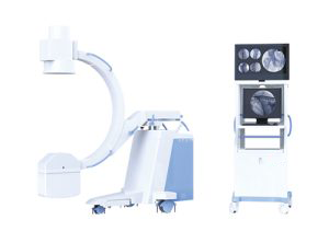
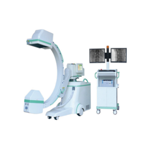
C700 is a highly reliable, tough, and enduring digital mobile system with a flat panel equipped with digital angiographic memory suitable for all the applications having high diagnostic content.
Features:
C700 (with Flat Panel) 30×30 or 21×21 cm
Digital Memories: 1,5k x 1,5k, with acquisition up to 25 fps (for Xenox C400);
30 kW H.V. Generator
DUAL COOLING System for immediate and effective heat removal DUAL POWER System: power reserve system
C-Arm ORBITAL Movement: 150° (105°, -45°); 150° (105°, -45°)
WIG-WAG: ± 10°; ± 10°
Useful space: 810 mm; 810 mm
C-Arm Depth: 640 mm; 640 mm
Min/ Max Height: 1455 / 1955 mm; 1455 / 1955 mm
Min/Max Length: 2085/ 2295 mm; 2110/ 2285 mm
Max Width: 800 mm; 800 mm
I.D.: 1065 mm; 1065 mm
Operating modes and functionalityOperating modalities of (rtp) memory
continuous fluoroscopy
high dose pulsed fluoroscopy
pulsed fluorography
digital snapshot
fluoroscopy ma : (range: 0,5-8 ma)
radiography: 2 points technique (kv and mas)
cine sequence: up to 25 fps. (included in basic configuration)
Software base funtion (live processing):
display of patient data, hospital name, x-ray parameters on operator monitor
recursive filter for noise reduction
dynamic recursive filter for patient’s movement compensation
edge enhancement spatial filter
edge enhancement spatial filter
horizontal and vertical image inversion
digital zoom during fluoroscopy mode (new)
metal detection in manual or automatic mode (new)
continuous digital image rotation
adjustable contrast and brightness
adaptive gamma curve
continuous fluoroscopy modality with lih (last image hold)
low dose fluoroscopy
timer function
measurements: length, angles, with manual calibration system, text overlay
anatomic program set up
80 anatomic programs in fluoroscope
Post-processing base features:
last image hold (lih)
last fluoroscopy run hold (lrh)
horizontal/vertical image inversion
digital image rotation
greyscale image inversion
adjustable contrast and brightness
adjustable gamma curve (out)
edge enhancement
electronic shutters (rectangular and circular)
electronic zoom roam from 1x to 5x
electronic mobile lens
mosaic overview
patient’s selection for exams and images management
patient’s exams transfer in the Dicom system
Applications:
Electrophysiology studies
Angiographic procedures
Vascular surgery
Endovascular applications
Urology procedures
Catheter procedures
Neuroradiology
General surgery
Traumatology
Orthopaedic
Interventional radiology
Options
DSA OPTION: Real time DSA with mask, Real time DSA with CO2 contrast liquid, Max opacity modality, Land marking, Road mapping, Mask selection for road mapping function, Virtual Road Mapping using fluorography image, Autoinjector delay selection, “CO2” contrast injection modality, Vascular tracing Function, Mask replace function, “Pixel shifting” function, Stenosimisure. In case the options are present all the relative functions are activated.
Dicom function (option): Dicom verify, Dicom storage, Dicom worklist, Dicom media interchange, Dicom send, Dicom mpps, Dicom structured report, Dicom query/retrieve (q/r), Dicom print interface with Dicom film editor




Reviews
There are no reviews yet.